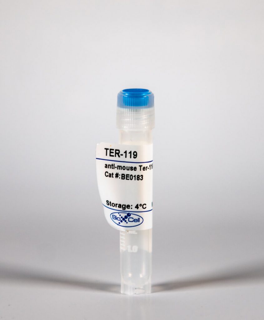
InVivoMab anti-mouse Ter-119
| Clone | TER-119 | ||||||||||||
|---|---|---|---|---|---|---|---|---|---|---|---|---|---|
| Catalog # | BE0183 | ||||||||||||
| Category | InVivoMab Antibodies | ||||||||||||
| Price |
|
The TER-119 monoclonal antibody reacts with mouse Ter-119 a 52 kDa glycophorin A-associated protein that is expressed by erythroid cells from the early proerythroblast stage to mature erythrocytes. The TER-119 antibody is commonly used for identifying erythrocytes and cells in the erythroid lineage.'
| Isotype | Rat IgG2b, κ |
| Recommended Isotype Control(s) | InVivoMAb rat IgG2b isotype control, anti-keyhole limpet hemocyanin(BE0090) |
| Recommended InVivoPure Dilution Buffer | InVivoPure pH 7.0 Dilution Buffer(IP0070) |
| Immunogen | C57BL/6 mouse fetal liver cells |
| Reported Applications |
|
| Endotoxin |
|
| Purity |
|
| Formulation |
|
| Sterility | 0.2 μM filtered |
| Production | Purified from tissue culture supernatant in an animal free facility |
| Purification | Protein G |
| Storage | The antibody solution should be stored at the stock concentration at 4°C. Do not freeze. |
| RRID | AB_10949625 |
| Molecular Weight | 150 kDa |
InVivoMAb anti-mouse Ter-119 (Clone: TER-119 )
Becker, A. M., et al. (2015). "ADAM17 limits the expression of CSF1R on murine hematopoietic progenitors." Exp Hematol 43(1): 44-52 e41-43. PubMed
All-lymphoid progenitors (ALPs) yield few myeloid cells in vivo, but readily generate such cells in vitro. The basis for this difference remains unknown. We hypothesized that ALPs limit responsiveness to in vivo concentrations of myeloid-promoting cytokines by reducing expression of the corresponding receptors, potentially through posttranscriptional mechanisms. Consistent with such a mechanism, ALPs express higher levels of CSF1R transcripts than their upstream precursors, yet show limited cell-surface protein expression of colony-stimulating factor 1 receptor (CSF1R). All-lymphoid progenitors and other hematopoietic progenitors deficient in A disintegrin and metalloproteinase domain 17 (ADAM17), display elevated cell surface CSF1R expression. ADAM17(-/-) ALPs, however, fail to yield myeloid cells upon transplantation into irradiated recipients. Moreover, ADAM17(-/-) ALPs yield fewer macrophages in vitro than control ALPs at high concentrations of macrophage colony stimulating factor. Mice with hematopoietic-specific deletion of ADAM17 have normal numbers of myeloid and lymphoid progenitors and mature cells in vivo. These data demonstrate that ADAM17 limits CSF1R protein expression on hematopoietic progenitors, but that compensatory mechanisms prevent elevated CSF1R levels from altering lymphoid progenitor potential.
Yu, X., et al. (2015). "A monoclonal antibody with anti-D-like activity in murine immune thrombocytopenia requires Fc domain function for immune thrombocytopenia ameliorative effects." Transfusion 55(6 Pt 2): 1501-1511. PubMed
BACKGROUND: The mechanism of action of anti-D in ameliorating immune thrombocytopenia (ITP) remains unclear. The monoclonal antibody (MoAb) Ter119, which targets murine red blood cells (RBCs), has been shown to mimic the effect of anti-D in improving antibody-mediated murine ITP. The mechanism of Ter119-mediated ITP amelioration, especially the role of the antigen-binding and Fc domains, remains untested. A functional Fc domain is crucial for many therapeutic MoAb activity; therefore, the requirement of Ter119 Fc domain in ITP amelioration is investigated using outbred CD-1 mice. STUDY DESIGN AND METHODS: Ter119 variants, including Ter119 F(ab')2 fragments, deglycosylated Ter119, and afucosylated Ter119, were generated to test their effect in ameliorating antibody-induced murine ITP. In vivo inhibition of FcgammaRIII and FcgammaRIIB was achieved using the Fab fragment of the FcgammaRIII/FcgammaRIIB-specific MoAb 2.4G2. RESULTS: Ter119 F(ab')2 fragments and deglycosylated Ter119 were unable to ameliorate murine ITP or mediate phagocytosis of RBCs by RAW264.7 macrophages in vitro. Inhibition of FcgammaRIII and FcgammaRIIB, as well as Ter119 defucosylation, do not affect Ter119-mediated ITP amelioration. CONCLUSION: The Fc domain of Ter119, as well as its Fc glycosylation, is required for Ter119-mediated ITP amelioration. Moreover, both Fc and Fc glycosylation are required for Ter119-mediated phagocytosis in vitro. These findings demonstrate the importance of the Fc domain in a therapeutic MoAb with anti-D-like activity.
Fraser, S. T., et al. (2007). "Maturation and enucleation of primitive erythroblasts during mouse embryogenesis is accompanied by changes in cell-surface antigen expression." Blood 109(1): 343-352. PubMed
Primitive erythroblasts (EryPs) are the first hematopoietic cell type to form during mammalian embryogenesis and emerge within the blood islands of the yolk sac. Large, nucleated EryPs begin to circulate around midgestation, when connections between yolk sac and embryonic vasculature mature. Two to 3 days later, small cells of the definitive erythroid lineage (EryD) begin to differentiate within the fetal liver and rapidly outnumber EryPs in the circulation. The development and maturation of EryPs remain poorly defined. Our analysis of embryonic blood at different stages reveals a stepwise developmental progression within the EryP lineage from E9.5 to E12.5. Thereafter, EryDs are also present in the bloodstream, and the 2 lineages are not easily distinguished. We have generated a transgenic mouse line in which the human epsilon-globin gene promoter drives expression of green fluorescent protein exclusively within the EryP lineage. Here, we have used this line to characterize changes in cell morphology and surface-marker expression as EryPs mature and to track EryP numbers and enucleation throughout gestation. This study identifies previously unrecognized synchronous developmental stages leading to the maturation of EryPs in the mouse embryo. Unexpectedly, we find that EryPs are a stable cell population that persists through the end of gestation.
Otani, T., et al. (2004). "Erythroblasts derived in vitro from embryonic stem cells in the presence of erythropoietin do not express the TER-119 antigen." Exp Hematol 32(7): 607-613. PubMed
OBJECTIVE: In this study, we analyzed murine primitive erythropoiesis by coculturing Flk-1+ ES-derived cells with OP9 to find efficient culture conditions for erythroid cell induction. We utilized a nonserum culture system and EPO (erythropoietin) and found that this cytokine had unique properties. MATERIALS AND METHODS: ES cells (E14.1) were first differentiated to Flk-1+ cells and then cocultured with OP9 stromal cells. BIT9500 was used as a serum replacement. The erythroid morphology, hemoglobin types, and TER-119 expression levels were analyzed. RESULTS: Primitive erythroid cells with embryonic hemoglobin were generated very efficiently when the serum-containing culture was converted to the nonserum system. In this serum-free culture, TER-119+ erythroblasts appeared first on day 2 and maturation proceeded until day 7. When EPO was added to this coculture, the number of induced floating cells increased twofold to threefold. Unexpectedly, the erythroid-specific antigen TER-119 expression of these cells was drastically reduced. Since reduced TER-119 expression is usually interpreted as maturation arrest, we examined the phenotypic features of the EPO-treated cells. We found, however, no evidence of maturation arrest in the aspects of morphology and hemoglobin content. EPO did not suppress TER-119 expression of erythroblasts derived from fetal liver or adult bone marrow. CONCLUSIONS: Our results showed that EPO had the unusual property of inducing TER-119- erythroblasts in ES-derived primitive erythropoiesis. It is likely that this effect is unique to primitive erythropoiesis.


